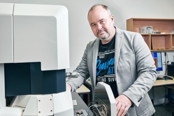
"Our laboratory is open not only for academics from our faculty, but basically for all students from CTU or other universities who need to measure their own samples and thus support their research results in the field of natural sciences or electronic components. It is also a great opportunity for students to get acquainted with the state-of-the-art facilities of contemporary microscopy. The new laboratory will thus contribute to providing the labour market with highly specialised experts, who are in demand particularly from companies operating in the fields of biomedicine, nanomaterials, electronic components, semiconductors or batteries," says Prof. Bohuslav Rezek, Head of the Department of Physics at FEL CTU, who leads the team of the Correlation Microscopy Laboratory.
Students address problems how to detect resistant bacteria and how to replace ineffective antibiotics
Presently, the laboratory is used by four research and academic staff from the department and a PhD student Ing. Markéta Šlapal Bařinková and Bc. Daniel Vítek, a master's student of Medical Electronics and Bioinformatics at FEL CTU, who are working on their projects here.
Markéta Šlapal Bařinková highlights the contribution of the new laboratory to the study of the interaction between bacteria and photoactive nanoparticles, a topic she is investigating in her PhD thesis. "The electron microscope makes it possible to find individual bacteria and distinguish between the different components of a sample based on electron contrast. The atomic force microscope then helps to characterize changes in 3D topography. The ability to prepare samples at any time and look at bacteria and their interactions with nanoparticles at the nanometer scale opens the door to obtaining new information and performing new analyses relatively easily," explains the PhD student from the Department of Physics. Her work aims to contribute to the detection of bacteria resistant to antimicrobial agents, one of the most intensively researched problems in medical science today.
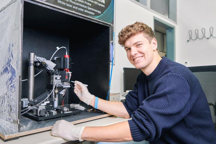
Daniel Vitek's thesis is also related to the search for alternatives to current drugs. In it, the student investigates electrochemical biosensors with nanodiamonds and gold nanoparticles for better detection of biomolecules and biomarkers. "In the long term, we would like to achieve improved detection of health conditions and drug effects using nanoparticles," says Daniel Vítek, who, like Markéta Šlapal Bařinková, could not imagine his research without the instrumentation of the new laboratory. "In the laboratory, with the help of my more experienced colleagues, I use a new AFM microscope, which provides me with valuable information on the topography, morphology or electrochemical properties of nanomaterials and confirms or refutes theories I have arrived at due to measurements on other instruments. At the same time, the microscope photographs are very attractive for planned publications," summarises the student, who admits that he has developed a considerable fondness for observing the micro and nano world.
The laboratory was established as part of an international project involving five teams from the Czech Republic and Korea
The reference laboratory will be involved in the demonstration, testing and further development of CPEM (Correlative Probe-Electron Microscopy) measurements. This method is developed by NenoVision and combines signals from an atomic force microscope (AFM) and a scanning electron microscope (SEM) into a final comprehensive data set and its image visualization correlating selected morphological, chemical, mechanical, electrical and magnetic properties. Applications of this method range from life sciences to semiconductor and nanomaterial analysis.
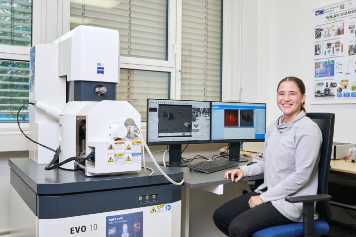
The cooperation with the Brno-based company Nenovision was crucial for the opening of the laboratory. The company was behind the development of a specialised AFM module, which the laboratory acquired as part of the cooperation on the TACOM joint project. The AFM module was developed specifically for integration into the SEM, where, with advanced electronic and software control, the sample under investigation can be imaged by both instruments simultaneously. The SEM microscope with appropriate parameters was supplied by Zeiss.
The reference laboratory was established at FEL in 2023 as part of the international TACOM project supported by the TAČR (Technology Agency of Czech Republic) grant agency. The project was launched in March last year and involves a total of five teams from the Czech Republic and Korea. While the Korean side focuses mainly on the development of the so-called AirSEM microscope for Life Sciences (a scanning electron microscope for the examination of biological samples outside the vacuum), the Czech side is working intensively on the research and development of correlative SEM-AFM microscopy. One of the objectives of the project is to strengthen research and industrial development cooperation between the Czech Republic and Korea to facilitate the development, integration and wider commercial application of these methods.
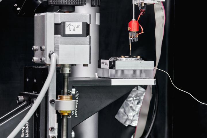
Within the TACOM project, Prof. Bohuslav Rezek's group at the FEL CTU Department of Physics collaborates on research and development of new methods of correlative microscopy. The reference laboratory is used for demonstration and testing of measurements in CPEM mode. Apart from this method, the laboratory also contains other instruments for correlative measurements, in particular a multifunctional optical microscope, which includes Raman spectrometer, a photoluminescence spectrometer, an atomic force microscope, Kelvin microscopy, electrical conductivity measurements and a scanning near-field microscope.
Illustrative photography can be found here. Source: Petr Neugebauer, FEL CTU